Rapid Weight Gain: An Unusual Presentation of Leukemia
CC BY-NC-ND 4.0 · Indian J Med Paediatr Oncol 2020; 41(02): 257-259
DOI: DOI: 10.4103/ijmpo.ijmpo_241_18
Abstract
Neurological manifestations of leukemia can be due to direct effects of the malignancy or due to the indirect effects of infection or therapy. An 11-year-old boy presented with recent-onset weight gain with papilledema and a history of tuberculosis contact. Neuroimaging and initial microscopic examination of the cerebrospinal fluid (CSF) did not aid in the diagnosis of central nervous system leukemia. He was started on antitubercular treatment yet deteriorated. Repeat CSF analysis when subjected to cytospin and flow cytometry confirmed the diagnosis of B cell acute lymphoblastic leukemia (ALL). Although there have been reports of relapsed ALL presenting as obesity, isolated rapid changes in weight at initial presentation are a very rare and unusual manifestation of ALL. To the best of our knowledge, this is the first such report.
Publication History
Received: 03 November 2018
Accepted: 17 September 2019
Article published online:
23 May 2021
© 2020. Indian Society of Medical and Paediatric Oncology. This is an open access article published by Thieme under the terms of the Creative Commons Attribution-NonDerivative-NonCommercial-License, permitting copying and reproduction so long as the original work is given appropriate credit. Contents may not be used for commercial purposes, or adapted, remixed, transformed or built upon. (https://creativecommons.org/licenses/by-nc-nd/4.0/.)
Thieme Medical and Scientific Publishers Pvt. Ltd.
A-12, 2nd Floor, Sector 2, Noida-201301 UP, India
Abstract
Neurological manifestations of leukemia can be due to direct effects of the malignancy or due to the indirect effects of infection or therapy. An 11-year-old boy presented with recent-onset weight gain with papilledema and a history of tuberculosis contact. Neuroimaging and initial microscopic examination of the cerebrospinal fluid (CSF) did not aid in the diagnosis of central nervous system leukemia. He was started on antitubercular treatment yet deteriorated. Repeat CSF analysis when subjected to cytospin and flow cytometry confirmed the diagnosis of B cell acute lymphoblastic leukemia (ALL). Although there have been reports of relapsed ALL presenting as obesity, isolated rapid changes in weight at initial presentation are a very rare and unusual manifestation of ALL. To the best of our knowledge, this is the first such report.
Keywords
Leukemia - meningitis - rapid weight gain - tuberculosisIntroduction
Acute leukemia is the most common childhood malignancy constituting 30% of all childhood malignancies with acute lymphoblastic leukemia (ALL) accounting for the majority.[1] In India, according to data obtained from the Population-based Cancer Registries, the age-adjusted incidence rates ranged from 0.0 pm/year (North East) to 101.4 pm/year (North) for boys and from 0.0 pm/year in West and North East to 62.3 pm/year in North region of India for girls.[2] Neurological complications of leukemia include intracranial hemorrhage, dural venous sinus thrombosis, seizures, focal neurological deficits, and cranial nerve palsies. Rarely, rapid weight gain can be the initial manifestation of childhood leukmia and must be differentiated from other causes of obesity.
Case Report
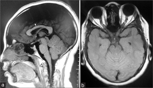
An 11-year-old boy was brought to our hospital with recent-onset weight gain of 3 months’ duration. His appetite was normal. He did not complain of headache, vomiting, seizures, or visual disturbances. His scholastic performance was satisfactory and there were no behavioral changes. His grandmother had passed away 4 years back due to pulmonary tuberculosis and his father had been recently diagnosed with pulmonary tuberculosis. At the time of admission, he weighed 45 kg with a body mass index (BMI) of 22 kg/m2 (between 1 and 2 standard deviation [SD] on the WHO BMI for age chart corresponding to the 97th percentile). His preillness weight was 35 kg with BMI 17. He was afebrile, with a pulse rate of 90 beats/min and the blood pressure (BP) recorded was 104/74 mmHg which was between the 50th and 90th spercentile for age and height. He had minimal right lateral rectus palsy and bilateral papilledema. The rest of his systemic examination was normal. Peripheral smear revealed a normocytic normochromic blood picture with no atypical cells. Chest radiograph was normal. Mantoux was negative. Other investigations including liver and renal function tests and lipid and thyroid profile were normal. His retroviral status was negative. Magnetic resonance imaging (MRI) of the brain showed sulcal hyperintensities suggestive of exudates with buckling of the optic nerve. The sella, pituitary, and parasellar structures were normal with normal ventricles [Figure 1]. His baseline investigations and cerebrospinal fluid (CSF) analysis are given in [Table 1]. However, due to lymphocytic meningitis and a history of contact with tuberculosis, he was started dexamethasone initially at 0.6 mg/kg/day followed by oral prednisolone at 60 mg/day and antitubercular drugs and discharged. He was advised to take full-dose steroids for 6 weeks followed by a slow taper over 2–4 weeks.

| Figure 1: Magnetic resonance imaging: (a) normal ventricles, sella, pituitary, parasellar structures. No macroadenoma (b) buckling of optic nerve
Table 1 Investigations
One month later, he had gained another 10 kg and complained of acute-onset diplopia. There was no fever, headache, vomiting, or seizures. He appeared cushingoid, with bilateral proptosis. He weighed 55 kg with a BMI of 24 kg/m2 (between 2 and 3 SD on the WHO BMI for age chart indicating he was at risk for obesity), BP was 140/110 mmHg (>99th centile which was 121/88 mm Hg), and pulse rate was 92 beats/min. He complained of neck stiffness, and fundus examination showed Grade I hypertensive retinopathy. Rest of the systemic examination was normal. Basic investigations along with CSF study were repeated, and the findings are enumerated in [Table 1]. Peripheral blood smear showed normocytic normochromic blood picture with no atypical cells. Neuroimaging revealed the same findings as the previous MRI.
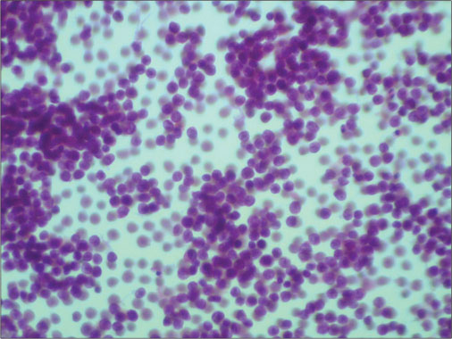
Cytospin performed on the CSF sample revealed a highly cellular monotonous population of uniform round cells with increased nuclear–cytoplasmic ratio, coarse chromatin, and indistinct nucleoli which were of leukemic origin [Figure 2]. Subsequently, flow cytometry and bone marrow study were done which confirmed B cell ALL.

| Figure 2: Highly cellular monotonous population of uniform round cells with increased nuclear-cytoplasmic ratio with coarse chromatin and indistinct nucleoli
This child was started on chemotherapy for high-risk ALL. However, after a period of 1 year, he succumbed to his illness.
Discussion
Neurological manifestations of leukemia can be due to direct infiltration by the tumor or due to immunosuppression therapy. Acute leukemia, especially ALL, is more likely to cause meningeal invasion than other forms of leukemia. Meningeal involvement in leukemia can occur throughout the course of the disease from as early as 2 days after diagnosis to as late as 6 years.[3] It may be due to leukemic meningitis, subarachnoid hemorrhage, and chemical (following intrathecal administration of chemotherapy) or infectious causes (bacterial or fungal).
The causes of weight gain in leukemia can be multifactorial. It can be related to the disease process or arise during or after completion of treatment. Weight gain can be due to an alteration in the lifestyle and reduced physical activity. It can also be caused by hypothalamic damage from cranial irradiation, chemotherapy-induced obesity, secondary growth hormone deficiency, and prolonged corticosteroid intake.[4]
Central nervous system leukemia (CNSL) itself can cause rapid weight gain and obesity by different mechanisms. Leukemic infiltration can cause dysfunction of the ventromedial hypothalamus, which integrates the peripheral neural and hormonal afferent signals of satiety and neuroendocrine efferents to balance energy storage versus expenditure. Any disturbance in this equilibrium results in hyperphagia and vagally mediated augmentation of insulin production leading to intractable weight gain and subsequent obesity. Another plausible mechanism is the destruction of the corticotropin-releasing hormone system, which results in the removal of the restraining influence on the pituitary function causing hypersecretion of adrenocorticotropic hormone, and thereby adrenocortical hypertrophy and Cushing’s disease.[5]
A positive CSF cytology is seen in the initial lumbar puncture in 50%–70% of cases of leukemia with leptomeningeal involvement. The presence of CSF lymphocytes of B cell lineage is highly suggestive of leukemic meningitis as cells of T cell lineage are more likely to be reactive. Flow cytometry is a more sensitive mode of investigation than cytomorphology for the detection of leukemic cells in the CSF. It also helps in differentiating leukemic infiltrates from reactive pleocytosis in those cases of doubtful cytomorphological identification.[6] [7]
MRI findings in CNSL include leptomeningeal enhancement of the brain, spinal cord, cauda equina, or subependymal areas, which extend into the sulci of the cerebrum or folia of the cerebellum. In patients with rare manifestations of CNSL, a combination of cytomorphology, flow cytometry, and cranial MRI is more useful to reach an accurate diagnosis.[8]
Obesity has been found to have several associations with leukemia, but most of these mechanisms are related to treatment. There are very few reports in English literature where abnormal weight gain was the initial presentation, and all have been reported for the diagnosis of relapsed disease.[5] We report the first case wherein rapid weight gain was the first and sole manifestation of ALL. Despite the strong history of contact with tuberculosis and the presence of lymphocytic meningitis, the absence of fever with weight gain as the predominant symptom should prompt one to investigate thoroughly for other causes before treating for tuberculosis, however common it may be.
Declaration of patient consent
The authors certify that they have obtained all appropriate patient consent forms. In the form the patient and parents have given their consent for his images and other clinical information to be reported in the journal. The patients understand that their names and initials will not be published and due efforts will be made to conceal their identity, but anonymity cannot be guaranteed.
Conflict of Interest
There are no conflicts of interest.
Acknowledgment
We would like to thank Dr. Jerome Joseph, Assistant Surgeon, Government Hospital, Kuzhithurai, Tamil Nadu for his contributions to this paper.
References
- Childhood central nervous system leukemia: Historical perspectives, current therapy, and acute neurological sequelae. Neuroradiology 2007; 49: 873-88
- Asthana S, Labani S, Mehrana S, Bakhshi S. Incidence of childhood leukaemia and lymphoma in India. Pediatr Hematol Oncol J 2018; 3: 115-20
- Chamberlain MC. Leukemia and the nervous system. Curr Oncol Rep 2005; 7: 66-73
- Iughetti L, Bruzzi P, Predieri B, Paolucci P. Obesity in patients with acute lymphoblastic leukemia in childhood. Ital J Pediatr 2012; 38: 4
- Zhang LD, Li YH, Ke ZY, Huang LB, Luo XQ. Obesity as the initial manifestation of central nervous system relapse of acute lymphoblastic leukemia: Case report and literature review. J Cancer Res Ther 2012; 8: 151-3
- Roma AA, Garcia A, Avagnina A, Rescia C, Elsner B. Lymphoid and myeloid neoplasms involving cerebrospinal fluid: Comparison of morphologic examination and immunophenotyping by flow cytometry. Diagn Cytopathol 2002; 27: 271-5
- Beatrice AM, Selvan C, Mukhopadhyay S. Pituitary dysfunction in infective brain diseases. Indian J Endocrinol Metab 2013; 17: S608-11
- Ulu EM, Töre HG, Bayrak A, Güngör D, Coşkun M. MRI of central nervous system abnormalities in childhood leukemia. Diagn Interv Radiol 2009; 15: 86-92
Address for correspondence
Publication History
Received: 03 November 2018
Accepted: 17 September 2019
Article published online:
23 May 2021
© 2020. Indian Society of Medical and Paediatric Oncology. This is an open access article published by Thieme under the terms of the Creative Commons Attribution-NonDerivative-NonCommercial-License, permitting copying and reproduction so long as the original work is given appropriate credit. Contents may not be used for commercial purposes, or adapted, remixed, transformed or built upon. (https://creativecommons.org/licenses/by-nc-nd/4.0/.)
Thieme Medical and Scientific Publishers Pvt. Ltd.
A-12, 2nd Floor, Sector 2, Noida-201301 UP, India

| Figure 1: Magnetic resonance imaging: (a) normal ventricles, sella, pituitary, parasellar structures. No macroadenoma (b) buckling of optic nerve

| Figure 2: Highly cellular monotonous population of uniform round cells with increased nuclear-cytoplasmic ratio with coarse chromatin and indistinct nucleoli
References
- Childhood central nervous system leukemia: Historical perspectives, current therapy, and acute neurological sequelae. Neuroradiology 2007; 49: 873-88
- Asthana S, Labani S, Mehrana S, Bakhshi S. Incidence of childhood leukaemia and lymphoma in India. Pediatr Hematol Oncol J 2018; 3: 115-20
- Chamberlain MC. Leukemia and the nervous system. Curr Oncol Rep 2005; 7: 66-73
- Iughetti L, Bruzzi P, Predieri B, Paolucci P. Obesity in patients with acute lymphoblastic leukemia in childhood. Ital J Pediatr 2012; 38: 4
- Zhang LD, Li YH, Ke ZY, Huang LB, Luo XQ. Obesity as the initial manifestation of central nervous system relapse of acute lymphoblastic leukemia: Case report and literature review. J Cancer Res Ther 2012; 8: 151-3
- Roma AA, Garcia A, Avagnina A, Rescia C, Elsner B. Lymphoid and myeloid neoplasms involving cerebrospinal fluid: Comparison of morphologic examination and immunophenotyping by flow cytometry. Diagn Cytopathol 2002; 27: 271-5
- Beatrice AM, Selvan C, Mukhopadhyay S. Pituitary dysfunction in infective brain diseases. Indian J Endocrinol Metab 2013; 17: S608-11
- Ulu EM, Töre HG, Bayrak A, Güngör D, Coşkun M. MRI of central nervous system abnormalities in childhood leukemia. Diagn Interv Radiol 2009; 15: 86-92


 PDF
PDF  Views
Views  Share
Share

