 PDF
PDF  Views
Views  Share
Share


Mediastinal ganglioneuroma: An incidentaloma of childhood
CC BY-NC-ND 4.0 · Indian J Med Paediatr Oncol 2013; 34(02): 130-131
DOI: DOI: 10.4103/0971-5851.116218
Abstract
Ganglioneuroma is a rare benign neurogenic tumor which represents the final maturation stage of neuroblast tumors. Here, we are discussing an interesting case of incidentally detected posterior mediastinal ganglioneuroma which should be kept in mind when dealing with any child with respiratory distress.
Keywords
Asymptomatic mediastinal mass - childm incidentaloma - ganglioneuroma - posterior mediastinal ganglioneuromaPublication History
Article published online:
20 July 2021
© 2013. Indian Society of Medical and Paediatric Oncology. This is an open access article published by Thieme under the terms of the Creative Commons Attribution-NonDerivative-NonCommercial-License, permitting copying and reproduction so long as the original work is given appropriate credit. Contents may not be used for commercial purposes, or adapted, remixed, transformed or built upon. (https://creativecommons.org/licenses/by-nc-nd/4.0/.)
Thieme Medical and Scientific Publishers Pvt. Ltd.
A-12, 2nd Floor, Sector 2, Noida-201301 UP, India
Abstract
Ganglioneuroma is a rare benign neurogenic tumor which represents the final maturation stage of neuroblast tumors. Here, we are discussing an interesting case of incidentally detected posterior mediastinal ganglioneuroma which should be kept in mind when dealing with any child with respiratory distress.
IMAGES IN MEDICAL AND PEDIATRIC ONCOLOGY
A 5-year-old boy was admitted to a district hospital with diagnosis of a right hydrocele. Routine blood and urine examination were normal. Patient never had any respiratory problems and was taken up for surgery, but at the time of induction of anesthesia, the patient developed severe respiratory distress, so operation was postponed. Later, chest X-ray done showed a dense homogenous opacity at left middle and lower zone, arising from the left paracardiac region [Figure 1]. So, he was referred to our institute.
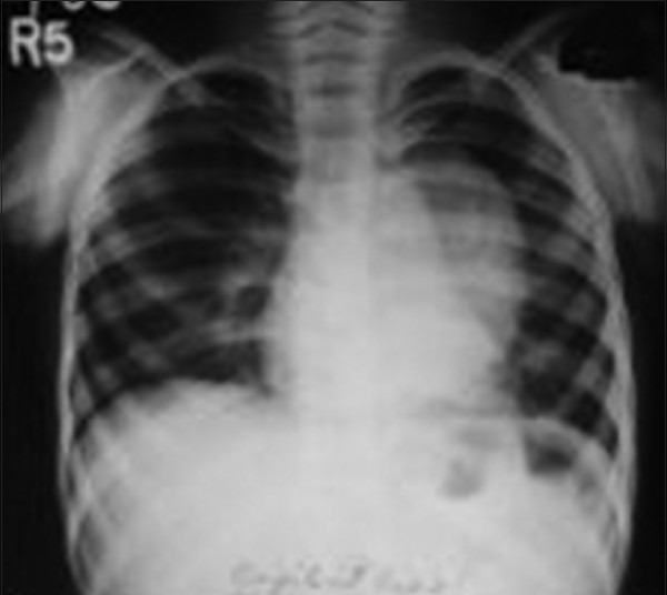
| Fig. 1 X-ray of thorax revealing large mass in left mediastinum
Computed tomography (CT) scan of the chest revealed a well-defined hypodense moderately enhancing non-calcified posterior mediastinal mass (4.9 × 4.6 × 6 cm3) in the left paravertebral region [Figure 2]. Magnetic resonance imaging (MRI) of the chest showed no extension of the tumor into the spinal canal [Figure 3]. Thus, excision of the mass was done by left posterior lateral thoracotomy [Figure 4]. Microscopically, the lesion was consistent with ganglioneuroma. Postoperative period was uneventful. Thirteen months after surgery, he is asymptomatic with normal radiology.
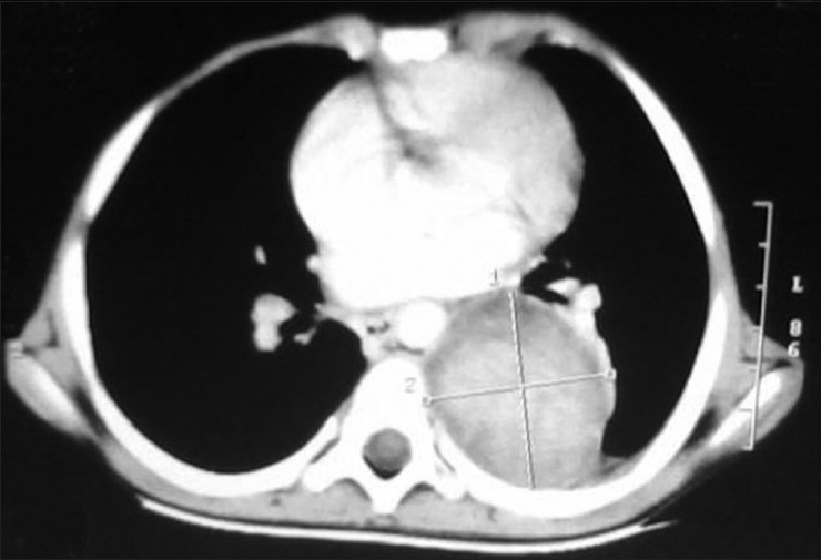
| Fig. 2 Computed tomography scan of thorax showing mass in the left posterior mediastinum region
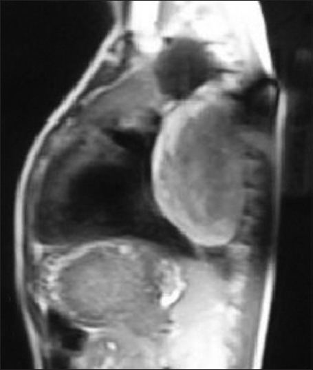
| Fig. 3 MRI of thorax showing posterior mediastinal mass with no spinal extension
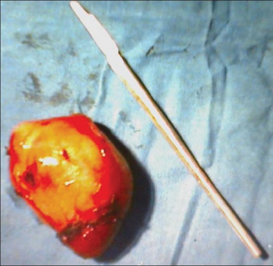
| Fig. 4 Excised specimen of posterior mediastinal mass
Footnotes
Source of Support: Nil
Conflict of Interest: None declared.
| Fig. 1 X-ray of thorax revealing large mass in left mediastinum
| Fig. 2 Computed tomography scan of thorax showing mass in the left posterior mediastinum region
| Fig. 3 MRI of thorax showing posterior mediastinal mass with no spinal extension
| Fig. 4 Excised specimen of posterior mediastinal mass