 PDF
PDF  Views
Views  Share
Share


Lacrimal gland enlargement in acute myeloid leukemia
CC BY-NC-ND 4.0 · Indian J Med Paediatr Oncol 2013; 34(01): 54-55
DOI: DOI: 10.4103/0971-5851.113441

Publication History
Article published online:
20 July 2021
© 2013. Indian Society of Medical and Paediatric Oncology. This is an open access article published by Thieme under the terms of the Creative Commons Attribution-NonDerivative-NonCommercial-License, permitting copying and reproduction so long as the original work is given appropriate credit. Contents may not be used for commercial purposes, or adapted, remixed, transformed or built upon. (https://creativecommons.org/licenses/by-nc-nd/4.0/.)
Thieme Medical and Scientific Publishers Pvt. Ltd.
A-12, 2nd Floor, Sector 2, Noida-201301 UP, India
Sir,
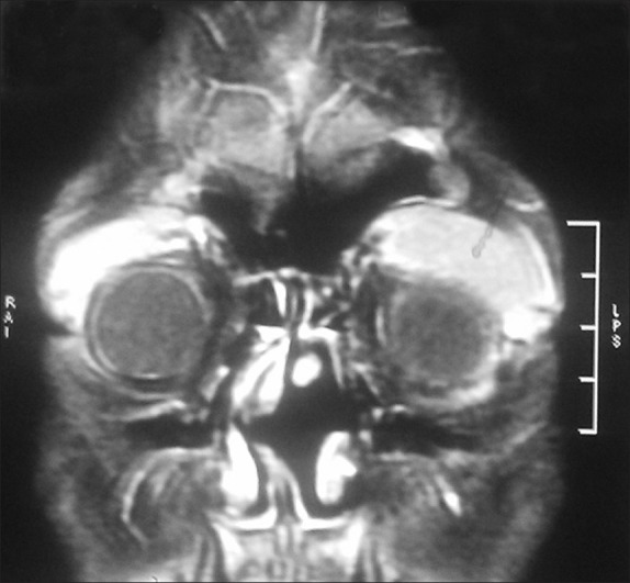
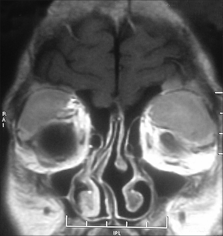
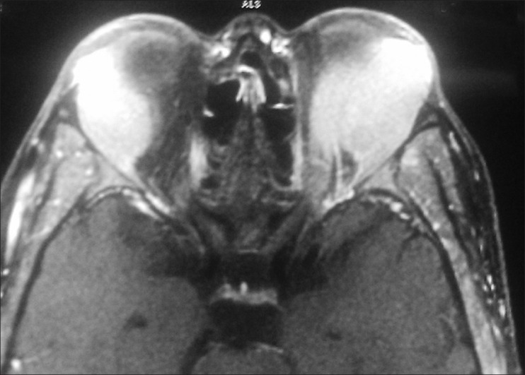
An 18-year-old male got admitted to emergency room (ER) of our hospital, a tertiary care center, for evaluation of bilateral protrusion of eyeballs. There was history of easy fatigability and ill-health for past 2 months. His clinical examination revealed anemia and bilateral proptosis. CBC showed Hb 8 g/dl, TLC 18 k, DLC 80% myeloblasts, platelets 70 k. Computed tomography (CT) scan of the orbits revealed lacrimal gland enlargement pushing eyeballs medially and down [Figures [Figures11–4]. The patient was shifted to medical oncology department on the same day.
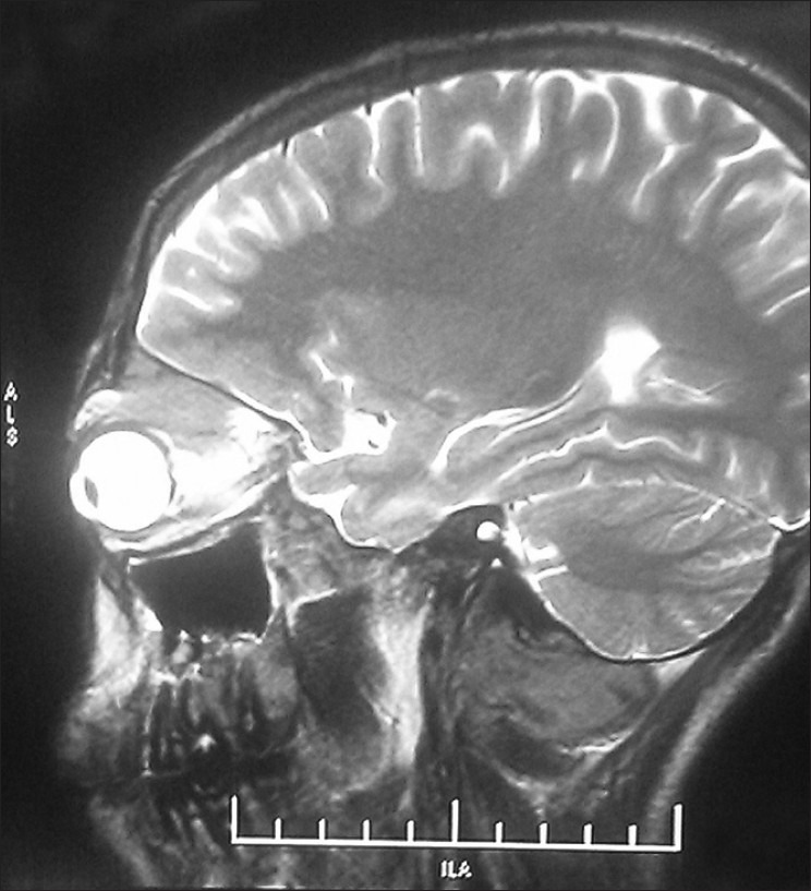
| Fig. 1 Eye balls pushed in CT orbit
| Fig. 4 Eyeballs compressed supero laterally
| Fig. 2 Lacrimal gland enlarged
| Fig. 3 Enlarged lacrimals pushing eyeballs
The aim of highlighting this image is how varied can be the differential diagnosis in a patient presenting as proptosis.
| Fig. 1 Eye balls pushed in CT orbit
| Fig. 4 Eyeballs compressed supero laterally
| Fig. 2 Lacrimal gland enlarged
| Fig. 3 Enlarged lacrimals pushing eyeballs