Giant mediastinal carcinoid
CC BY-NC-ND 4.0 · Indian J Med Paediatr Oncol 2015; 36(03): 194
DOI: DOI: 10.4103/0971-5851.166764

Publication History
Article published online:
12 July 2021
© 2015. Indian Society of Medical and Paediatric Oncology. This is an open access article published by Thieme under the terms of the Creative Commons Attribution-NonDerivative-NonCommercial-License, permitting copying and reproduction so long as the original work is given appropriate credit. Contents may not be used for commercial purposes, or adapted, remixed, transformed or built upon. (https://creativecommons.org/licenses/by-nc-nd/4.0/.)
Thieme Medical and Scientific Publishers Pvt. Ltd.
A-12, 2nd Floor, Sector 2, Noida-201301 UP, India
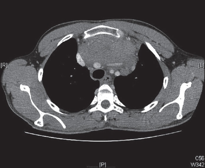
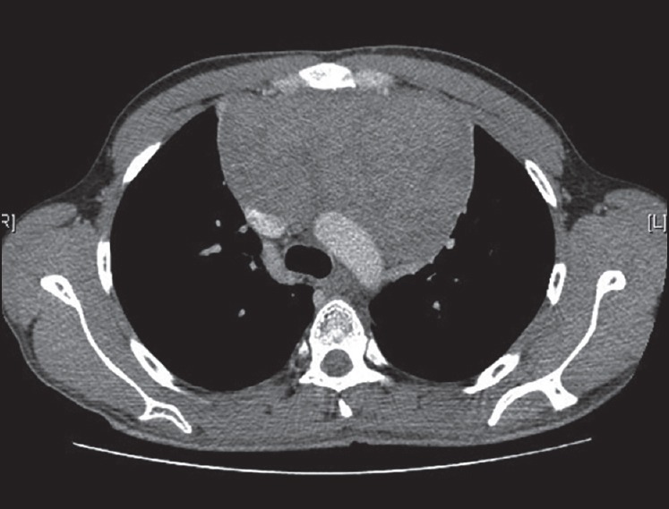
A 25-year-old male was presented with cough. Computed tomography (CT) scan showed anterior mediastinal mass [Figure 1] encasing the aortic arch and left brachiocephalic vein with compression of the superior vena cava. CT-guided biopsy showed neuroendocrine tumor staining with chromogranin, synaptophysin, and neuron specific enolase. Postchemoradiotherapy positron emission tomography-CT scan revealed static disease [Figure 2]. At surgery, the mass could be dissected off the major vessels; the thrombosed left brachiocephalic vein was resected. Good collaterals obviated the need for vascular reconstruction. The patient recovered after a stormy postoperative course. Histopathology report showed neuroendocrine carcinoma, mitotic rate of 10/10 HPF, and MIB-1 proliferation index of 8-10%. On 5-year follow-up, the patient is asymptomatic and disease-free on CT scan.

| Fig. 1 Anterior mediastinal mass encasing arch aorta, left brachiocephalic vein, and compressing superior vena cava

| Fig. 2 Static disease post chemoradiotherapy

| Fig. 1 Anterior mediastinal mass encasing arch aorta, left brachiocephalic vein, and compressing superior vena cava

| Fig. 2 Static disease post chemoradiotherapy


 PDF
PDF  Views
Views  Share
Share

