Ewing’s Sarcoma of the Vulva: An Uncommon Tumor in an Uncommon Site
CC BY-NC-ND 4.0 · Indian J Med Paediatr Oncol 2020; 41(03): 397-399
DOI: DOI: 10.4103/ijmpo.ijmpo_57_20
Introduction
Extraskeletal Ewing's sarcoma (EES) is an uncommon, aggressive, round cell tumor. It accounts to 16% among Ewing's sarcoma (ES). The most commonly involved sites are soft tissues of trunk or lower extremities, paravertebral tissues, chest wall, and retroperitoneum. EES presenting in the genitourinary tract is highly uncommon. There are very few cases of primary vulvar and vaginal ES which have been published to date. We report a rare case of ES of the vulva that presented with labial swelling.
Publication History
Received: 12 February 2020
Accepted: 01 May 2020
Article published online:
28 June 2021
© 2020. Indian Society of Medical and Paediatric Oncology. This is an open access article published by Thieme under the terms of the Creative Commons Attribution-NonDerivative-NonCommercial-License, permitting copying and reproduction so long as the original work is given appropriate credit. Contents may not be used for commercial purposes, or adapted, remixed, transformed or built upon. (https://creativecommons.org/licenses/by-nc-nd/4.0/.)
Thieme Medical and Scientific Publishers Pvt. Ltd.
A-12, 2nd Floor, Sector 2, Noida-201301 UP, India
Introduction
Extraskeletal Ewing's sarcoma (EES) is an uncommon, aggressive, round cell tumor. It accounts to 16% among Ewing's sarcoma (ES). The most commonly involved sites are soft tissues of trunk or lower extremities, paravertebral tissues, chest wall, and retroperitoneum. EES presenting in the genitourinary tract is highly uncommon. There are very few cases of primary vulvar and vaginal ES which have been published to date. We report a rare case of ES of the vulva that presented with labial swelling.
Case Presentation
A 53-year-old postmenopausal female with no comorbidities presented with left labial swelling for 5 months. She was evaluated elsewhere and found to have a 3 cm × 3 cm left labial swelling. She underwent excision biopsy of the lesion and was referred to us for further management.
Question 1
Clinical differential diagnosis of such presentation in this age group?
Answer
-
Vulvar cysts (Bartholin's cyst)
-
Cutaneous adnexal tumors
-
Carcinoma of the vulva
-
Melanoma.
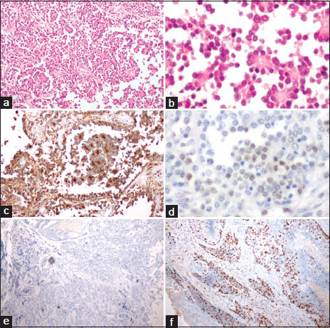
Histopathology showed small round blue cells arranged in diffuse sheets intervened by fibrous septae. The cells had minimal cytoplasm, vesicular nuclear chromatin, and inconspicuous nucleoli forming rosettes at places. Mitosis was brisk with areas of necrosis, suggestive of malignant round cell tumor of the left labia. Immunohistochemistry was positive for CD99 and Friend leukemia virus integration 1 (FLI-1) with focal synaptophysin positivity. Ki67 index was 20%. Features were suggestive of ES. Fluoresence in situ hybridisation (FISH) for ES breakpoint region 1 (EWSR1) gene rearrangement was done. Tumor cells were positive for EWSR1 gene translocation (t [22q12]) [Figure 1].

| Figure 1:Histopathology of the resected specimen (a and b) low‑power (H and E × 100) and high‑power view (H and E × 400), respectively, showing sheets of small round blue cells forming rosettes at places; (c and d) immunohistochemistry showing neoplastic cells positive for CD99 and Friend leukemia virus integration 1, respectively; (e) immunohistochemistry showing neoplastic cells negative for leukocyte common antigen (f) Ki67 index of 20%.
Question 2
Percentage of ES with EWS-FLI1?
Answer
t (11; 22) (q24; q12) leading to a chimeric transcript EWS-FLI1 is seen in 85% of cases. In 5%–10%, EWS gene is fused with other members of Erythroblast Transformation-Specific (ETS) genes like ETS-related gene (ERG), ETS variant transcription factor 1 (ETV1), ETS variant transcription factor 4 (ETV4) and Fifth Ewing variant (FEV).[1]
Contrast-enhanced computed tomography of the thorax, abdomen, and pelvis was done and did not show any residual disease, but was suggestive of postoperative changes in the left labia and subcentimetric retroperitoneal and pelvic nodes. Bone scan did not show any tracer uptake in the bones. Bone marrow aspiration and biopsy from the right iliac crest did not show any involvement by the tumor. A diagnosis of ES of Vulva pT1NoMo G3 Stage II was made.
Question 3
Incidence of ES of vulva?
Answer
EES of the female genital tract (FGT) is very rare. The most common sites in FGT were the ovaries followed by uterine body, cervix, vagina, and vulva.[2] Less than 30 cases of ES of the vulva have been reported till date. To the best of our knowledge, we report the 5th case in the country.
The patient was started on VAC/IE (injection vincristine 1.5 mg/m2, injection adriamycin 75 mg/m2, injection cyclophosphamide 1.2 g/m2 (D1)/injection ifosfamide 1.8 g/m2, and injection etoposide 100 mg/m2 (D1–D5) regimen). She has received four cycles of chemotherapy with resolution of iliac lymph nodes on contrast-enhanced computed tomography. She is planned for a total of 17 cycles of chemotherapy.
Discussion
ES is a highly malignant, small, round cell tumor which originates from primitive neuroectodermal cells first described by James Ewing in 1921.[3] It is predominantly seen in males with a male-to-female ratio of 1.5:1.[4] In general, 20%–30% of patients present with metastases at the time of their diagnosis.[5]
Extraskeletal Ewing's sarcoma (EES) is uncommon. It accounts to 16% among ES.[6] They usually occur in the soft tissue of trunk, lower extremities, paravertebral tissues, chest wall, and retroperitoneum. A few reported rare sites include larynx, nasal cavity, neck, lung, retroperitoneum, mediastinum, and genital tract. EES of the FGT is very rare, and the reported most common sites in FGT were the ovaries[7] followed by the uterine body[8] and less commonly the cervix,[9] vagina, and vulva.[2],[10] EES usually occur in relatively young women; the mean age at the time of the initial diagnosis of previous cases is 27.6 years (median: 26, range: 10–52 years),[11] but our patient was older than this.
The differential diagnosis of these tumors includes other small round blue cell tumors such as rhabdomyosarcoma, lymphoma, small-cell carcinoma (primary or metastatic), melanoma, cutaneous adnexal tumors, and Merkel cell carcinoma.
Demonstration of t(11;22) (q24;q12) chromosomal translocation (EWS-FLI1 gene rearrangement) is highly specific for ES/primitive neuroectodermal tumors since it is seen in more than 90% of the tumors.
The treatment of ES requires a multidisciplinary approach. It includes surgical resection with multiagent chemotherapy. Radiation therapy may be used to improve local control in cases of R1/R2 resection. The multiagent standard chemotherapy regimen typically includes doxorubicin, vincristine, and cyclophosphamide, alternating with ifosfamide and etoposide.[12] Doxorubicin is replaced with actinomycin-D once the cumulative dose of 375 mg/m2 is crossed.
EES has more aggressive behavior and a worse prognosis than their bony counterpart. Reported 5-year survival rate for skeletal ES is around 75% as compared to 38% in extraskeletal counterpart. However, there is favorable outcome for extraosseous ES arising in cutaneous and other superficial site.[13],[14] In the absence of metastatic disease, primary vulvar and vaginal ES neoplasms likely have a more favorable prognosis. Hence, prognosis depends on tumor size, superficial location, early detection, metastasis, and complete surgical removal of the lesion.[15] According to literature in metastatic cases, most patients either died of disease or were alive with disease during their last follow-up. In nonmetastatic lesions, there was survival benefit ranging from 18 to 51 months.[16]
Conclusion
Early diagnosis and multimodality treatment are necessary for survival benefit. Because of fewer reported cases in literature, it is difficult to draw any conclusion about tumor behavior, epidemiology, or standard management. Hence, studies of more number of cases of primary vulvar ES with longer follow-up periods are needed to clarify the same.
Declaration of patient consent
The authors certify that they have obtained all appropriate patient consent forms. In the form, the patient has given her consent for her images and other clinical information to be reported in the journal. The patient understands that name and initials will not be published and due efforts will be made to conceal identity, but anonymity cannot be guaranteed.
Acknowledgment
Residents and faculty of Department of Pathology and Medical Oncology
Conflict of Interest
There are no conflicts of interest.
References
- Rekhi B, Chinnaswamy G, Vora T, Shah S, Rangarajan V. Primary Ewing sarcoma of vulva, confirmed with molecular cytogenetic analysis: A rare case report with diagnostic and treatment implications. Indian J Pathol Microbiol 2015; 58: 341-4
- Yang J, Guo Q, Yang Y, Zhang J, Lang J, Shi H. Primary vulvar Ewing sarcoma/primitive neuroectodermal tumor: A report of one case and review of the literature. J Pediatr Adolesc Gynecol 2012; 25: e93-7
- Ewing J. Diffuse endothelioma of bone. Pathol Soc 1921; 21: 17-24
- Yeshvanth SK, Ninan K, Bhandary SK, Lakshinarayana KP, Shetty JK, Makannavar JH. Rare case of extra skeletal Ewings sarcoma of the sinonasal tract. J Cancer Res Ther 2012; 8: 142-4
- Cotterill SJ, Ahrens S, Paulussen M, Jürgens HF, Voûte PA, Gadner H. et al. Prognostic factors in Ewing's tumor of bone: Analysis of 975 patients from the European Intergroup Cooperative Ewing's Sarcoma Study Group. J Clin Oncol 2000; 18: 3108-14
- Pradhan A, Grimer RJ, Spooner D, Peake D, Carter SR, Tillman RM. et al. Oncological outcomes of patients with Ewing's sarcoma: Is there a difference between skeletal and extra-skeletal Ewing's sarcoma?. J Bone Joint Surg B 2011; 93: 531-6
- Kleinman GM, Young RH, Scully RE. Primary neuroectodermal tumors of the ovary. A report of 25 cases. Am J Surg Pathol 1993; 17: 764-78
- Snijders-Keilholz A, Ewing P, Seynaeve C, Burger CW. Primitive neuroectodermal tumor of the cervix uteri: A case report-changing concepts in therapy. Gynecol Oncol 2005; 98: 516-9
- Li B, Ouyang L, Han X, Zhou Y, Tong X, Zhang S. et al. Primary primitive neuroectodermal tumor of the cervix. Onco Targets Ther 2013; 6: 707-11
- Xiao C, Zhao J, Guo P, Wang D, Zhao D, Ren T. et al. Clinical analysis of primary primitive neuroectodermal tumors in the female genital tract. Int J Gynecol Cancer 2014; 24: 404-9
- Che SM, Cao PL, Chen HW, Liu Z, Meng D. Primary Ewing's sarcoma of vulva: A case report and a review of the literature. J Obstet Gynaecol Res 2013; 39: 746-9
- Grier HE, Krailo MD, Tarbell NJ, Link MP, Fryer CJ, Pritchard DJ. et al. Addition of ifosfamide and etoposide to standard chemotherapy for Ewing's sarcoma and primitive neuroectodermal tumor of bone. N Engl J Med 2003; 348: 694
- Al-Tamimi H, Al-Hadi AA, Al-Khater AH, Al-Bozom I, Al-Sayed N. Extra skeletal neuroectodermal tumour of the vagina: A single case report and review. Arch Gynecol Obstet 2009; 280: 465-8
- Tunitsky-Bitton E, Uy-Kroh MJ, Michener C, Tarr ME. Primary Ewing sarcoma presenting as a vulvar mass in an adolescent: Case report and review of literature. J Pediatr Adolesc Gynecol 2015; 28: e179-83
- Fong YE, López-Terrada D, Zhai QJ. Primary Ewing sarcoma/peripheral primitive neuroectodermal tumor of the vulva. Hum Pathol 2008; 39: 1535-9
- McCluggage WG, Sumathi VP, Nucci MR, Hirsch M, Dal Cin P, Wells M. et al. Ewing family of tumours involving the vulva and vagina: Report of a series of four cases. J Clin Pathol 2007; 60: 674-80
Address for correspondence
Publication History
Received: 12 February 2020
Accepted: 01 May 2020
Article published online:
28 June
2021
© 2020. Indian Society of Medical and Paediatric Oncology. This is an open access article published by Thieme under the terms of the Creative Commons Attribution-NonDerivative-NonCommercial-License, permitting copying and reproduction so long as the original work is given appropriate credit. Contents may not be used for commercial purposes, or adapted, remixed, transformed or built upon. (https://creativecommons.org/licenses/by-nc-nd/4.0/.)
Thieme Medical and Scientific Publishers Pvt. Ltd.
A-12, 2nd Floor, Sector 2, Noida-201301 UP,
India

| Figure 1:Histopathology of the resected specimen (a and b) low‑power (H and E × 100) and high‑power view (H and E × 400), respectively, showing sheets of small round blue cells forming rosettes at places; (c and d) immunohistochemistry showing neoplastic cells positive for CD99 and Friend leukemia virus integration 1, respectively; (e) immunohistochemistry showing neoplastic cells negative for leukocyte common antigen (f) Ki67 index of 20%
References
- Rekhi B, Chinnaswamy G, Vora T, Shah S, Rangarajan V. Primary Ewing sarcoma of vulva, confirmed with molecular cytogenetic analysis: A rare case report with diagnostic and treatment implications. Indian J Pathol Microbiol 2015; 58: 341-4
- Yang J, Guo Q, Yang Y, Zhang J, Lang J, Shi H. Primary vulvar Ewing sarcoma/primitive neuroectodermal tumor: A report of one case and review of the literature. J Pediatr Adolesc Gynecol 2012; 25: e93-7
- Ewing J. Diffuse endothelioma of bone. Pathol Soc 1921; 21: 17-24
- Yeshvanth SK, Ninan K, Bhandary SK, Lakshinarayana KP, Shetty JK, Makannavar JH. Rare case of extra skeletal Ewings sarcoma of the sinonasal tract. J Cancer Res Ther 2012; 8: 142-4
- Cotterill SJ, Ahrens S, Paulussen M, Jürgens HF, Voûte PA, Gadner H. et al. Prognostic factors in Ewing's tumor of bone: Analysis of 975 patients from the European Intergroup Cooperative Ewing's Sarcoma Study Group. J Clin Oncol 2000; 18: 3108-14
- Pradhan A, Grimer RJ, Spooner D, Peake D, Carter SR, Tillman RM. et al. Oncological outcomes of patients with Ewing's sarcoma: Is there a difference between skeletal and extra-skeletal Ewing's sarcoma?. J Bone Joint Surg B 2011; 93: 531-6
- Kleinman GM, Young RH, Scully RE. Primary neuroectodermal tumors of the ovary. A report of 25 cases. Am J Surg Pathol 1993; 17: 764-78
- Snijders-Keilholz A, Ewing P, Seynaeve C, Burger CW. Primitive neuroectodermal tumor of the cervix uteri: A case report-changing concepts in therapy. Gynecol Oncol 2005; 98: 516-9
- Li B, Ouyang L, Han X, Zhou Y, Tong X, Zhang S. et al. Primary primitive neuroectodermal tumor of the cervix. Onco Targets Ther 2013; 6: 707-11
- Xiao C, Zhao J, Guo P, Wang D, Zhao D, Ren T. et al. Clinical analysis of primary primitive neuroectodermal tumors in the female genital tract. Int J Gynecol Cancer 2014; 24: 404-9
- Che SM, Cao PL, Chen HW, Liu Z, Meng D. Primary Ewing's sarcoma of vulva: A case report and a review of the literature. J Obstet Gynaecol Res 2013; 39: 746-9
- Grier HE, Krailo MD, Tarbell NJ, Link MP, Fryer CJ, Pritchard DJ. et al. Addition of ifosfamide and etoposide to standard chemotherapy for Ewing's sarcoma and primitive neuroectodermal tumor of bone. N Engl J Med 2003; 348: 694
- Al-Tamimi H, Al-Hadi AA, Al-Khater AH, Al-Bozom I, Al-Sayed N. Extra skeletal neuroectodermal tumour of the vagina: A single case report and review. Arch Gynecol Obstet 2009; 280: 465-8
- Tunitsky-Bitton E, Uy-Kroh MJ, Michener C, Tarr ME. Primary Ewing sarcoma presenting as a vulvar mass in an adolescent: Case report and review of literature. J Pediatr Adolesc Gynecol 2015; 28: e179-83
- Fong YE, López-Terrada D, Zhai QJ. Primary Ewing sarcoma/peripheral primitive neuroectodermal tumor of the vulva. Hum Pathol 2008; 39: 1535-9
- McCluggage WG, Sumathi VP, Nucci MR, Hirsch M, Dal Cin P, Wells M. et al. Ewing family of tumours involving the vulva and vagina: Report of a series of four cases. J Clin Pathol 2007; 60: 674-80


 PDF
PDF  Views
Views  Share
Share

