Detection and Significance of Human Epidermal Growth Factor Receptor 2 Expression in Gastric Adenocarcinoma
CC BY-NC-ND 4.0 · Indian J Med Paediatr Oncol 2017; 38(02): 153-157
DOI: DOI: 10.4103/ijmpo.ijmpo_159_16
Abstract
Background: Human epidermal growth factor receptor 2 (HER2) is involved in the pathogenesis of several types of cancer, including gastric cancer. Overexpression of HER2 is noted in 10%–22.8% of gastric adenocarcinoma and its identification is of immense importance for management by targeted drugs. Detection of HER2 expression in gastric malignancies has not been undertaken previously in the local population. Objective: To ascertain HER2 immunohistochemical expression in gastric adenocarcinoma and its relationship with the anatomic location and histomorphology. Materials and Methods: A total of 47 cases of gastric adenocarcinoma diagnosed over 2 years constituted the study group. Clinical history, type of operation, gross morphology, and hematoxylin and eosin (H and E) stained sections were reviewed. Two paraffin blocks were selected, immunostain was performed using rabbit monoclonal HER2 antibody and Hoffmann scoring system was applied. Results: Most of gastric carcinomas occurred in male (42 cases), having a mean age of 53.6 years. A total of eight cases (17.1%) had expressed a score of 3+ HER2 positivity. All positivity was noted in intestinal type according to Lauren classification (25%) and none in diffuse type. All HER2 score of 3+ was noted in histological grade of well and moderately differentiated adenocarcinoma. Score 2+ was noted in seven cases, among them, only two were poorly differentiated gastric adenocarcinoma. Conclusion: HER2 overexpression was noticeably associated with an intestinal subtype, and well and moderately differentiated adenocarcinomas. Such cases of gastric adenocarcinoma are considered for targeted therapy with trastuzumab in the local population.
Keywords
Gastric adenocarcinoma - histological grade - human epidermal growth factor receptor 2 expression - human epidermal growth factor receptor 2 scorePublication History
Article published online:
06 July 2021
© 2017. Indian Society of Medical and Paediatric Oncology. This is an open access article published by Thieme under the terms of the Creative Commons Attribution-NonDerivative-NonCommercial-License, permitting copying and reproduction so long as the original work is given appropriate credit. Contents may not be used for commercial purposes, or adapted, remixed, transformed or built upon. (https://creativecommons.org/licenses/by-nc-nd/4.0/.)
Thieme Medical and Scientific Publishers Pvt. Ltd.
A-12, 2nd Floor, Sector 2, Noida-201301 UP, India
Abstract
Background:
Human epidermal growth factor receptor 2 (HER2) is involved in the pathogenesis of several types of cancer, including gastric cancer. Overexpression of HER2 is noted in 10%–22.8% of gastric adenocarcinoma and its identification is of immense importance for management by targeted drugs. Detection of HER2 expression in gastric malignancies has not been undertaken previously in the local population.
Objective:
To ascertain HER2 immunohistochemical expression in gastric adenocarcinoma and its relationship with the anatomic location and histomorphology.
Materials and Methods:
A total of 47 cases of gastric adenocarcinoma diagnosed over 2 years constituted the study group. Clinical history, type of operation, gross morphology, and hematoxylin and eosin (H and E) stained sections were reviewed. Two paraffin blocks were selected, immunostain was performed using rabbit monoclonal HER2 antibody and Hoffmann scoring system was applied.
Results:
Most of gastric carcinomas occurred in male (42 cases), having a mean age of 53.6 years. A total of eight cases (17.1%) had expressed a score of 3+ HER2 positivity. All positivity was noted in intestinal type according to Lauren classification (25%) and none in diffuse type. All HER2 score of 3+ was noted in histological grade of well and moderately differentiated adenocarcinoma. Score 2+ was noted in seven cases, among them, only two were poorly differentiated gastric adenocarcinoma.
Conclusion:
HER2 overexpression was noticeably associated with an intestinal subtype, and well and moderately differentiated adenocarcinomas. Such cases of gastric adenocarcinoma are considered for targeted therapy with trastuzumab in the local population.
Introduction
Gastric cancer is the fourth most common malignancy worldwide and the second most common cause of cancer-related death. It is often diagnosed at a late stage, and there is a high frequency of metastasis, making therapy unsatisfactory. A number of chemotherapeutic agents have conventionally been used. Despite treatment, patients have a mean survival of <1 href="https://www.ncbi.nlm.nih.gov/pmc/articles/PMC5582552/#ref1" rid="ref1" class=" bibr popnode tag_hotlink tag_tooltip" id="__tag_635949650" role="button" aria-expanded="false" aria-haspopup="true" xss=removed>1] Hence, there is an urgent need to identify molecular characteristics of gastric cancer pathogenesis, and thus to develop targeted therapies designed on a molecular level.
Human epidermal growth factor receptor 2 (HER2) is a tyrosine kinase and a member of the epidermal growth factor receptor family.[1] HER2 is a proto-oncogene encoded by ERBB2 on chromosome 17. HER2 is found in many tissues,[2] including breast, the gastrointestinal tract, kidney, and liver. In general, it suppresses apoptosis and promotes cellular proliferation, thus facilitating uncontrolled cell growth, leading to tumorigenesis.[3] HER2 acts as an oncogene in a wide range of cancers. It is now well accepted that HER2 status is of supreme importance in breast cancers, with positivity for HER2 indicating a poor prognosis.[4] HER2 studies have been carried out extensively in breast cancer but not in gastric cancer. With increasing understanding of the molecular biology of HER2 and the availability of genomics and proteomics analyses, it has now been recognized that HER2 is implicated in other severe forms of cancer, notably gastric cancer.[5] Unlike the consensus in breast cancer, results of different studies in gastric cancer[6] have yielded inconsistent findings regarding prognosis, some studies have shown a worse prognosis[7] whereas others found no association.[8]
Trastuzumab, a recombinant humanized monoclonal antibody that targets the extracellular domain IV of HER2, is now a well-established modality of treatment in breast cancer. Trastuzumab has demonstrated a survival advantage in patients with HER2-overexpressed gastric cancer in few recent studies.[9]
A few studies have correlated HER2 overexpression with adenocarcinoma variants and anatomical location of gastric carcinoma. However, no studies have been done among local population for overexpression of HER2 and its morphological variants.
The present observational study was undertaken to determine HER2 status in patients with gastric adenocarcinoma and its correlation with clinical features, anatomic location, histological variants of gastric adenocarcinoma, grade, and stage of the disease.
Materials and Methods
This study is an observational, retrospective, and prospective study. The study population were cases of histopathologically diagnosed gastric adenocarcinoma, over 2 years. Patients having gastric carcinoma of other histopathological types were excluded from the study. After seeking the permission from the Institutional Ethical Committee, following data were obtained in each case of the study group – clinical history, type of surgical procedure, anatomic location, and gross morphology.
For the histopathological study, specimens were fixed in 10% neutral buffered formalin. Tissues were processed, embedded in paraffin, sections were taken and stained with H and E. After histopathological diagnosis of adenocarcinoma, cases were classified according to Lauren classification and tumor, node and metastasis (TNM) staging of each case was noted. Slides were reviewed and two blocks for each case were selected for immunohistochemistry. Respective paraffin blocks were retrieved, 4 μm sections were taken and mounted on poly-L lysine coated slides taking care to mount the tissue sections flat and wrinkle-free. Slides were incubated for 30 min at 60° centigrade. Slides were deparaffinized and rehydrated in graded alcohol and then in water. Antigen retrieval was done using Tris-ethylene-diamine-tetraacetic acid buffer at pH 9 in a domestic microwave in three cycles, for about 20 min. After cooling down to room temperature, slides were washed in wash buffer (pH 7.4). Endogenous peroxidase blocking was done with 3% hydrogen peroxide for 10 min. Immunohistochemistry was done using primary antibody (rabbit monoclonal HER2 antibody, clone SP3, Cell Marque) and slides were incubated for 60 min at room temperature in a moist chamber. Again after wash with wash buffer slides were incubated in the moist chamber with secondary antibody (horseradish peroxidase labeled). The reaction product was detected with 3,3-diaminobenzidinechromogen. Nuclear staining was carried out with Hematoxylin. Sections were again dehydrated in graded alcohol and mounted in dibutyl pthalate xylene.
Immunohistochemistry slides were scored by two pathologists individually according to the scoring system by Hofmann et al.[10] HER2 and fluorescent in situ hybridization positive breast cancer tissue was used as external positive control. All cases with score 3+ were considered positive.
Results
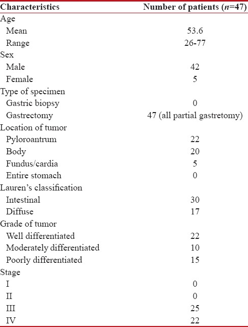
A total of 47 patients were included in the study group who fulfilled the inclusion criteria. A partial gastrectomy was done in all cases and histopathologically diagnosed to be gastric adenocarcinoma. Clinicopathological characteristics are depicted in Table 1. The age range of the study group was 26–77 years with mean age 53.6 years. Majority of patients were male, sex ratio (male:female) being 8.4:1
Table 1
Clinicopathological characteristics of patients
|
Al specimens were of complete or partial distal gastrectomy. Tumors were located at the cardiac end (5 cases, 10.6%), body (20 cases, 42.6%), and pyloroantrum (22 cases, 46.8%). Most of the lesions were grossly ulcerated, except 17 cases (36.2%) where the whole thickness of the stomach wall was diffusely involved.
Routine histopathological examination with H and E staining revealed gastric adenocarcinoma and were grouped according to Lauren classification. Intestinal type of adenocarcinoma [Figure 1] comprised 30/47 cases (63.8%), whereas the diffuse type of adenocarcinoma [Figure 2] was diagnosed in 17/47 cases (36.2%) [Table 2].
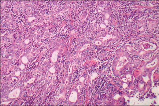
| Figure 1:Well-differentiated adenocarcinoma of intestinal type (H and E, ×100)
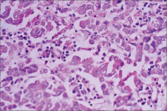
| Figure 2:Poorly differentiated adenocarcinoma (H and E, ×400)
Table 2
Human epidermal growth factor receptor 2 positivity according to Lauren's classification

|
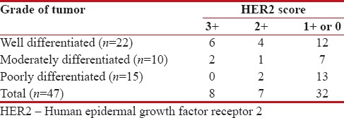
Regarding grading of tumors, well (22 cases), and moderately (10 cases) differentiated adenocarcinoma was noted in 32/47 patients (68.1%) and poorly differentiated adenocarcinoma accounted for 15/47 cases (31.9%) [Table 3].
Table 3
Human epidermal growth factor receptor 2 positivity according to tumor grade
|
With respect to staging, 25 patients (53.2%) had Stage III disease and 22 patients (46.8%) had Stage IV disease.
Immunohistochemistry for HER2/neu showed a score of 3+ positivity [Figure 3] in 8 cases (17.1%), all were adenocarcinoma of intestinal variant having well and moderate differentiation [Tables [Tables22 and and3]3] and originated from the body and pyloroantrum (four each). HER2 score was 2+ [Figure 4] in 7 cases (14.9%) these were again well to moderately differentiated adenocarcinoma of the intestinal variant. HER2 score was 1+ or 0 [Figure 5] in 32 cases (68.1%). In this group, 13 cases (40.6%) were graded as poorly differentiated, and among them eight cases were diffuse variant of adenocarcinoma. Only two cases of poorly differentiated, one of moderately differentiated carcinoma of intestinal variant and four cases of well-differentiated carcinoma showed 2+ HER2 positivity. Well and moderately differentiated carcinoma showed more HER2 3+ positivity (8 cases, 25%), whereas cases of poorly differentiated carcinoma predominantly (86.7%) showed the lack of HER2 expression. HER2 positivity score of 2+ was noted in seven cases of Stage III disease, whereas score 3+ was noted four cases each of Stage III and Stage IV disease, respectively. HER2 score was analyzed according to the grade of adenocarcinoma [Table 4]. A score of 2+ or 3+ was noted in 13 (40.6%) cases of well and moderately differentiated carcinoma but none in poorly differentiated carcinoma.
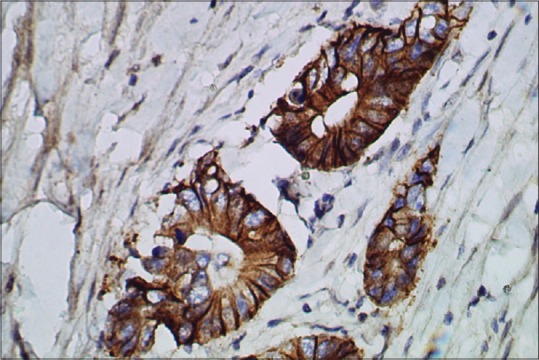
| Figure 3:Well-differentiated adenocarcinoma showing human epidermal growth factor receptor 2 score of 3+ (human epidermal growth factor receptor 2 immunostain, ×400)
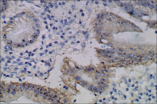
| Figure 4:Well-differentiated adenocarcinoma showing human epidermal growth factor receptor 2 score of 2+ (human epidermal growth factor receptor 2 immunostain, ×400)
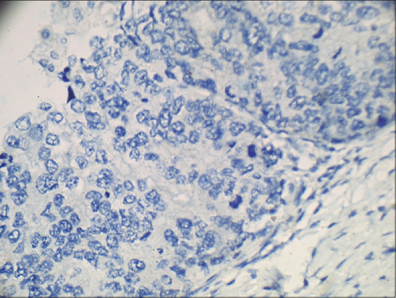
| Figure 5:Poorly differentiated adenocarcinoma showing negative human epidermal growth factor receptor 2 staining (human epidermal growth factor receptor 2 score of 0) (human epidermal growth factor receptor 2 immunostain ×400)

|
Discussion
Worldwide prevalence rates of HER2 expression in gastric adenocarcinomas ranges from 10% to 22.8%.[11] In a few study conducted in South Indian population, the expression is higher, varying from 35.89% to 44.2%.-[12,13] However, even after an extensive search of the literature, no data were available in the Eastern region of India. The present study, conducted among the patients from different Districts of West Bengal, showed the occurrence of HER2/neu 3+ expression in 8/47 cases (17.1%).
The present study had detected HER2 positivity in four tumors each situated at the pyloroantrum and body of the stomach. De Carli et al. found 6/7 (85.7%) HER2/neu positivity in pyloric antrum.[11] Lakshmi et al. found pyloroantrum positivity in 51.2%-cases.[12]
HER2 is more expressed in the intestinal type than diffuse type of gastric adenocarcinomas. Prevalence in intestinal type ranging from 6.1%-to 28.57%, and in the diffuse type from 0.7%-to 13.43%.[11] This strong correlation is supported by the present study with 8-out of 30-cases (26.7%) of the intestinal type of gastric adenocarcinomas showing HER2 positivity.
The present study found 25% HER2 positivity in well-differentiated adenocarcinomas. Lakshmi et al. in a study from South India found 60% HER2 positivity more in well-differentiated adenocarcinoma which is statistically significant, compared to 29.1%, in moderately differentiated adenocarcinoma and 20.6% in poorly differentiated adenocarcinoma. Ling et al. also found statistically significant correlation of HER2/neu positivity in well or moderately differentiated adenocarcinomas.[12,14]
Several studies concluded that there is no correlation between HER2 overexpression and pT, N, or M factors or TNM stages.[14,15,16] The present study also did not find any correlation with TNM staging. Qiu et al. proposed Lauren classification combined with HER2 status as a better prognostic factor than Lauren classification alone.[17]
Recently published data, from the randomized, prospective Phase III clinical trial ToGA provided first documentation of the clinical benefit of trastuzumab when used in combination with chemotherapy in the setting of advanced gastric and gastroesophageal junction cancer. Median overall survival was 13.8 months in trastuzumab arm compared to 11.1 months in the chemotherapy only arm. As a result of the survival benefit, it has become necessary to test the HER2 status and determine patient eligibility for targeted therapy.[18]
Conclusion
An accurate assessment of HER2 expression in gastric cancer patients is of immense importance and utility in the optimal selection of patients for trastusumab (Herceptin) therapy. The present study is reporting a significant HER2 overexpression of 16% in gastric adenocarcinomas. The present study also found that HER2 overexpression was more in intestinal type and in well and moderately differentiated gastric adenocarcinoma suggesting that these may be the candidates for targeted therapy using Herceptin. Larger studies and clinical trials are suggested to establish the these observations.
Financial support and sponsorship
Nil
Conflicts of interest
There are no conflicts of interest.
References
- Fornaro L, Lucchesi M, Caparello C, Vasile E, Caponi S, Ginocchi L, et al. Anti-HER agents in gastric cancer: From bench to bedside. Nat Rev Gastroenterol Hepatol 2011;8:369-83.
- Coussens L, Yang-Feng TL, Liao YC, Chen E, Gray A, McGrath J, et al. Tyrosine kinase receptor with extensive homology to EGF receptor shares chromosomal location with neu oncogene. Science 1985;230:1132-9.
- Neve RM, Lane HA, Hynes NE. The role of overexpressed HER2 in transformation. Ann Oncol 2001;12 Suppl 1:S9-13.
- Slamon DJ, Clark GM, Wong SG, Levin WJ, Ullrich A, McGuire WL. Human breast cancer: Correlation of relapse and survival with amplification of the HER-2/neu oncogene. Science 1987;235:177-82.
- Fukushige S, Matsubara K, Yoshida M, Sasaki M, Suzuki T, Semba K, et al. Localization of a novel v-erbB-related gene, c-erbB-2, on human chromosome 17 and its amplification in a gastric cancer cell line. Mol Cell Biol 1986;6:955-8.
- Gómez-Martin C, Garralda E, Echarri MJ, Ballesteros A, Arcediano A, RodrÁguez-Peralto JL, et al. HER2/neu testing for anti-HER2-based therapies in patients with unresectable and/or metastatic gastric cancer. J Clin Pathol 2012;65:751-7.
- Jørgensen JT, Hersom M. HER2 as a prognostic marker in gastric cancer – A systematic analysis of data from the literature. J Cancer 2012;3:137-44.
- Janjigian YY, Werner D, Pauligk C, Steinmetz K, Kelsen DP, Jäger E, et al. Prognosis of metastatic gastric and gastroesophageal junction cancer by HER2 status: A European and USA International collaborative analysis. Ann Oncol 2012;23:2656-62.
- Gunturu KS, Woo Y, Beaubier N, Remotti HE, Saif MW. Gastric cancer and trastuzumab:First biologic therapy in gastric cancer. Ther Adv Med Oncol 2013;5:143-51.
- He C, Bian XY, Ni XZ, Shen DP, Shen YY, Liu H, et al. Correlation of human epidermal growth factor receptor 2 expression with clinicopathological characteristics and prognosis in gastric cancer. World J Gastroenterol 2013;19:2171-8.
- De Carli DM, Rocha MP, Antunes LC, Fagundes RB. Immunohistochemical expression of HER2 in adenocarcinoma of the stomach. Arq Gastroenterol 2015;52:152-5.
- Lakshmi V, Valluru VR, Madhavi J, Valluru N. Role of her 2 neu in gastric carcinoma-3 year study in a medical college hospital. Indian J Appl Res 2014;4:1-4.
- Sekaran A, Kandagaddala RS, Darisetty S, Lakhtakia S, Ayyagari S, Rao GV, et al. HER2 expression in gastric cancer in Indian population – An immunohistochemistry and fluorescence in situ hybridization study. Indian J Gastroenterol 2012;31:106-10.
- Ling S, Jianming Y, Ning L. Her 2 expression and relevant clinicopathological features & gastroesophageal junction adenocarcinoma in a Chinese population. Diagn Pathol 2013;8:76.
- Kunz PL, Mojtahed A, Fisher GA, Ford JM, Chang DT, Balise RR, et al. HER2 expression in gastric and gastroesophageal junction adenocarcinoma in a US population: Clinicopathologic analysis with proposed approach to HER2 assessment. Appl Immunohistochem Mol Morphol 2012;20:13-24.
- Kataoka Y, Okabe H, Yoshizawa A, Minamiguchi S, Yoshimura K, Haga H, et al. HER2 expression and its clinicopathological features in resectable gastric cancer. Gastric Cancer 2013;16:84-93.
- Qiu M, Zhou Y, Zhang X, Wang Z, Wang F, Shao J, et al. Lauren classification combined with HER2 status is a better prognostic factor in Chinese gastric cancer patients. BMC Cancer 2014;14:823.
- Kang Y, Van Cutsem E, Chung H, Shen L, Sawaki A, Lordick F, et al. Efficacy results from the ToGA trial: A phase III study of trastuzumab added to standard chemotherapy (CT) in first- line human epidermal growth factor receptor 2 (HER2)-positive advanced gastric cancer (GC). J Clin Oncol 27:18s, 2009 (Suppl; abstr LBA4509).

| Figure 1:Well-differentiated adenocarcinoma of intestinal type (H and E, ×100)

| Figure 2:Poorly differentiated adenocarcinoma (H and E, ×400)

| Figure 3:Well-differentiated adenocarcinoma showing human epidermal growth factor receptor 2 score of 3+ (human epidermal growth factor receptor 2 immunostain, ×400)

| Figure 4:Well-differentiated adenocarcinoma showing human epidermal growth factor receptor 2 score of 2+ (human epidermal growth factor receptor 2 immunostain, ×400)

| Figure 5:Poorly differentiated adenocarcinoma showing negative human epidermal growth factor receptor 2 staining (human epidermal growth factor receptor 2 score of 0) (human epidermal growth factor receptor 2 immunostain ×400)
References
- Fornaro L, Lucchesi M, Caparello C, Vasile E, Caponi S, Ginocchi L, et al. Anti-HER agents in gastric cancer: From bench to bedside. Nat Rev Gastroenterol Hepatol 2011;8:369-83.
- Coussens L, Yang-Feng TL, Liao YC, Chen E, Gray A, McGrath J, et al. Tyrosine kinase receptor with extensive homology to EGF receptor shares chromosomal location with neu oncogene. Science 1985;230:1132-9.
- Neve RM, Lane HA, Hynes NE. The role of overexpressed HER2 in transformation. Ann Oncol 2001;12 Suppl 1:S9-13.
- Slamon DJ, Clark GM, Wong SG, Levin WJ, Ullrich A, McGuire WL. Human breast cancer: Correlation of relapse and survival with amplification of the HER-2/neu oncogene. Science 1987;235:177-82.
- Fukushige S, Matsubara K, Yoshida M, Sasaki M, Suzuki T, Semba K, et al. Localization of a novel v-erbB-related gene, c-erbB-2, on human chromosome 17 and its amplification in a gastric cancer cell line. Mol Cell Biol 1986;6:955-8.
- Gómez-Martin C, Garralda E, Echarri MJ, Ballesteros A, Arcediano A, RodrÁguez-Peralto JL, et al. HER2/neu testing for anti-HER2-based therapies in patients with unresectable and/or metastatic gastric cancer. J Clin Pathol 2012;65:751-7.
- Jørgensen JT, Hersom M. HER2 as a prognostic marker in gastric cancer – A systematic analysis of data from the literature. J Cancer 2012;3:137-44.
- Janjigian YY, Werner D, Pauligk C, Steinmetz K, Kelsen DP, Jäger E, et al. Prognosis of metastatic gastric and gastroesophageal junction cancer by HER2 status: A European and USA International collaborative analysis. Ann Oncol 2012;23:2656-62.
- Gunturu KS, Woo Y, Beaubier N, Remotti HE, Saif MW. Gastric cancer and trastuzumab:First biologic therapy in gastric cancer. Ther Adv Med Oncol 2013;5:143-51.
- He C, Bian XY, Ni XZ, Shen DP, Shen YY, Liu H, et al. Correlation of human epidermal growth factor receptor 2 expression with clinicopathological characteristics and prognosis in gastric cancer. World J Gastroenterol 2013;19:2171-8.
- De Carli DM, Rocha MP, Antunes LC, Fagundes RB. Immunohistochemical expression of HER2 in adenocarcinoma of the stomach. Arq Gastroenterol 2015;52:152-5.
- Lakshmi V, Valluru VR, Madhavi J, Valluru N. Role of her 2 neu in gastric carcinoma-3 year study in a medical college hospital. Indian J Appl Res 2014;4:1-4.
- Sekaran A, Kandagaddala RS, Darisetty S, Lakhtakia S, Ayyagari S, Rao GV, et al. HER2 expression in gastric cancer in Indian population – An immunohistochemistry and fluorescence in situ hybridization study. Indian J Gastroenterol 2012;31:106-10.
- Ling S, Jianming Y, Ning L. Her 2 expression and relevant clinicopathological features & gastroesophageal junction adenocarcinoma in a Chinese population. Diagn Pathol 2013;8:76.
- Kunz PL, Mojtahed A, Fisher GA, Ford JM, Chang DT, Balise RR, et al. HER2 expression in gastric and gastroesophageal junction adenocarcinoma in a US population: Clinicopathologic analysis with proposed approach to HER2 assessment. Appl Immunohistochem Mol Morphol 2012;20:13-24.
- Kataoka Y, Okabe H, Yoshizawa A, Minamiguchi S, Yoshimura K, Haga H, et al. HER2 expression and its clinicopathological features in resectable gastric cancer. Gastric Cancer 2013;16:84-93.
- Qiu M, Zhou Y, Zhang X, Wang Z, Wang F, Shao J, et al. Lauren classification combined with HER2 status is a better prognostic factor in Chinese gastric cancer patients. BMC Cancer 2014;14:823.
- Kang Y, Van Cutsem E, Chung H, Shen L, Sawaki A, Lordick F, et al. Efficacy results from the ToGA trial: A phase III study of trastuzumab added to standard chemotherapy (CT) in first- line human epidermal growth factor receptor 2 (HER2)-positive advanced gastric cancer (GC). J Clin Oncol 27:18s, 2009 (Suppl; abstr LBA4509).


 PDF
PDF  Views
Views  Share
Share

